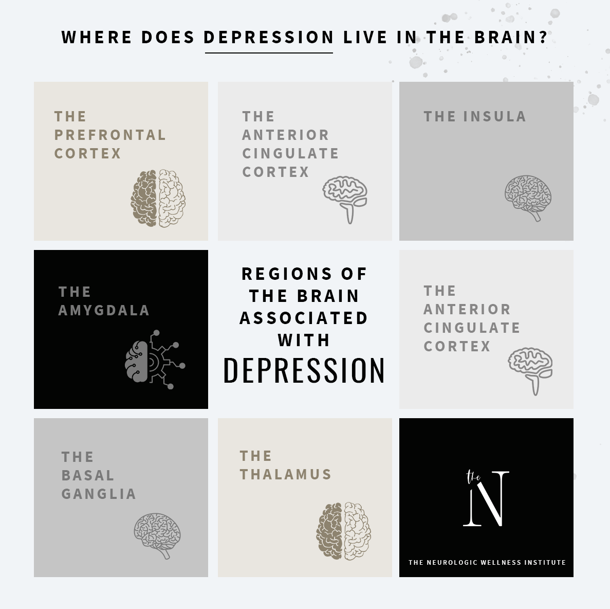
There have been multiple studies conducted attempting to study the areas of the brain involved in depression. Studies look at depression in the brain from various perspectives-functional, chemical, structural. For the purpose of this blog, we are going to focus on the structures in the brain that are involved in depression. Considering the many variations and causes of depression, each case may be different. However, despite the origin, there are certain areas in the brain that are directly or indirectly affected by and contribute to depression. Let’s take a closer look at these regions…..
The dorsolateral and dorsoventral regions of the prefrontal cortex
The role of the prefrontal cortex in depression may be altered through both structure and function. If there is not enough stimulation, fuel, oxygen, and circulation throughout the frontal region of the brain a depression in function may result. There have also been findings of decreased volume of the prefrontal cortex in those experiencing depression in which the size of that region of the brain has decreased.
The Insula
The insula is a region of the brain found in the cortex that separates the temporal lobe from the frontal and parietal lobes. It plays a major role in experiencing emotions such as negative emotions, disgust, internal body awareness, and response to various senses such as taste and smell. The activity of the insula increases during depression and is exceptionally sensitive to anything with a negative connotation.
The Limbic System
The limbic system is the region of the brain that regulates and responds to emotion and memory. It plays a major role in controlling how our hormones work, how our heart beats, how we breathe, and other physiological processes that are automatic for our bodies. Just as the limbic system is highly involved in anxiety, it also plays a major role in depression. To reiterate, limbic system is also composed of structures called the anterior cingulate cortex amygdala, the hippocampus, the basal ganglia, and the thalamus.
The Anterior Cingulate Cortex
The Anterior Cingulate Cortex plays a role in emotion and cognition. The front of the anterior cingulate cortex plays a role in communication within our limbic system or emotional regulating system while the back of the cortex plays a role in conflict resolution associated with things that create various types of emotion. It is the area of the brain that connects sight and smell with memory. Ever smell something and it instantly brings back a memory- good or bad? It is also the region of the brain that connects emotion and pain. The anterior cingulate cortex communicates with the hypothalamus in the brain which regulates our hormones, heart rate, breathing, and other bodily functions that we do not need to think about for them to occur. This is why people that suffer from depression tend to have problems with digestion, diarrhea, constipation, and hormonal abnormalities.
The Amygdala
The amygdala is a small structure located in the temporal lobes and is used in analyzing different emotions that arise in various situations. It increases our stress response if something is frightening or it creates new habits or fears based on the associated reward or punishment. It lies fairly close to the hippocampus and uses the memory of emotion to help form new memories–associating them as good or bad. With depression, negative emotions are often used as fuel in forming new memories leaving little room to form happy memories or create pleasant experiences. Recent studies have found that the size of the amygdala is relatively smaller in those with depression.
The Hippocampus
The Hippocampus’s primary role is to form new memories from past experiences. These memories are usually full of emotion.This area of the brain has also been found to be small in volume in those suffering from depression.
The Basal Ganglia
The basal ganglia is considered to be a part of the limbic system, but it is also involved in motor actions and responses-initiating and stopping movement. It plays a major role in learning new habits, responding to reward and motivation, forming sequencing actions, and memory formation. With depression, the constant negative reinforcement to the basal ganglia only creates a habitual response of negativity to the limbic circuit, ie. depression.
The Thalamus
The thalamus is the structure of the brain through which all sensory information needs to go through before being dispersed to the appropriate relay stations in the brain. Any type of stimulation involving emotion must pass through the thalamus. Generally, depression is a problem when the thalamus has difficulty activating and making the appropriate connections to other areas of the limbic system and the cortex. The “mood circuit” becomes reinforced with negativity and does not relay the appropriate messages back and forth.
It All Works Together
When it comes to depression, the structures listed above may individually contribute to depression. However, more than likely, it is the emotional response and communication between all of these structures that is the biggest contributing factor. Restructuring the circuit from a functional standpoint by providing the appropriate neurological reinforcement should ideally help to retrain the parts of the brain involved in emotional regulation.
Give us a call if you or a loved one is suffering from depression.
References:
Pandya, Mayur, et al. “Where in the Brain Is Depression?” Current Psychiatry Reports, vol. 14, no. 6, Nov. 2012, pp. 634–642., doi:10.1007/s11920-012-0322-7.
Boundless. “Boundless Psychology.” Lumen, https://courses.lumenlearning.com/boundless-psychology/chapter/structure-and-function-of-the-brain/.
